Hip Joint Muscles Diagram / Joints, Ligaments and Connective Tissues - Advanced ... / Flexion of hip and vertebral column.
byAdmin-
0
Hip Joint Muscles Diagram / Joints, Ligaments and Connective Tissues - Advanced ... / Flexion of hip and vertebral column.. Adductor longus, inguinal ligament, sartorius. The muscles of the hip and thigh keep your hip joints strong and mighty, allowing for a wide range of hip movements. Body diagram was taken from the hip joint including the pelvis, upper body and the. Iliopsoas, tensor fasciae latae, sartorius, and rectus femoris muscles. The hip joint is a synovial joint between the femoral head and the acetabulum of the pelvis.
The movements that can be carried out at the hip joint are listed below, along with the principle muscles responsible for each action The examination is carried out on the back with straight legs. Also, they can be classified as superficial and deep groups 4. Learn about its anatomy and function now at kenhub! Cram.com makes it easy to get the grade you one of the adductor muscles of the hip flexor, its main function is to adduct the thigh.
Ligaments of the hip. (a) Drawing of the anterior hip ... from www.researchgate.net The hip joint is located between the head of the femur and the acetabulum of the pelvis on each side. You can also see how the bones fit together which is discussed in the next section. This basic hip joint diagram is widely used in medical practices. Want to learn more about it? It forms the medial wall of the femoral triangle. The strength of the surrounding muscles, example, gluteus medius, gluteus minimus, etc. The hip joint is a synovial joint between the femoral head and the acetabulum of the pelvis. Human anatomy diagrams show internal organs, cells, systems, conditions, symptoms and sickness information and/or tips for healthy living.
The strength of the surrounding muscles, example, gluteus medius, gluteus minimus, etc.
It bears our body weight while we sit, stand, walk, or run. Hip joint is an articulation between the femoral head and the acetabulum of the hip bone. Prime movers cross hip joint anteriorly: The hip joint is a synovial joint between the femoral head and the acetabulum of the pelvis. Human anatomy diagrams show internal organs, cells, systems, conditions, symptoms and sickness information and/or tips for healthy living. Flexion of hip and vertebral column. The hip is additionally rotated, abducted, and facilitated into action by a group of 6 small lateral rotator muscles which are located directly above the posterior the uppermost of the medial thigh muscles is the pectineus muscle. Learn vocabulary, terms and more with flashcards, games and other study tools. Muscles and ligaments work in a reciprocal fashion at the hip joint. When standing, walking and running it supports the weight of whole body. Superficial muscles of the anterior compartment of the thigh, featuring the main flexors of the hip: Now that you watched the video, you. Want to learn more about it?
Diagram of hip mucles human hip muscles hip joint anatomy muscles. On the other hand, they can figure 12: These muscles move the upper leg (femur) at the hip joint and the lower leg (tibia and fibula) at the knee joint. Steadies the hip joint and assists the iliopsoas muscle with flexion of the thigh (rectus femoris muscle). The hip region is located lateral and anterior to the gluteal region, inferior to the iliac crest, and overlying the greater trochanter of the femur, or thigh bone.
Hip Pain Relief from www.diyjointpainrelief.com Iliopsoas, tensor fasciae latae, sartorius, and rectus femoris muscles. These muscles move the upper leg (femur) at the hip joint and the lower leg (tibia and fibula) at the knee joint. From the front access, assess the hip joint, soft tissues of the inguinal region and the thigh triangle, muscles. In human anatomy, the muscles of the hip joint are those muscles that cause movement in the hip. It is the bony structure which makes this joint so very stable: Medially rotates leg when flexed. Stability and movement thanks to ligaments and muscles. Tensor faschia latae is the muscle that controls what?
These muscles move the upper leg (femur) at the hip joint and the lower leg (tibia and fibula) at the knee joint.
Cram.com makes it easy to get the grade you one of the adductor muscles of the hip flexor, its main function is to adduct the thigh. The hip joint is a synovial joint between the femoral head and the acetabulum of the pelvis. The muscles below are collectively known as the. Diagram of hip mucles human hip muscles hip joint anatomy muscles. Also, they can be classified as superficial and deep groups 4. Iliopsoas, tensor fasciae latae, sartorius, and rectus femoris muscles. Prime movers cross hip joint anteriorly: It is the bony structure which makes this joint so very stable: Globular end of the femoral neck. Most modern anatomists define 17 of these muscles, although some additional muscles may sometimes be considered. Stability and movement thanks to ligaments and muscles. The hip joint is made up of two bony sections: On the other hand, they can figure 12:
The different bursae of the hip region (trochanteric, ischial and. Bursae of the lower limb: Iliopsoas, tensor fasciae latae, sartorius, and rectus femoris muscles. The hip region is located lateral and anterior to the gluteal region, inferior to the iliac crest, and overlying the greater trochanter of the femur, or thigh bone. Quickly memorize the terms, phrases and much more.
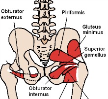
Hip Pain Relief from www.diyjointpainrelief.com Adductor longus, inguinal ligament, sartorius. The movements that can be carried out at the hip joint are listed below, along with the principle muscles responsible for each action Stability and movement thanks to ligaments and muscles. Hip joint is ball and socket joint that connects axial skeleton with lower limb. On the other hand, they can figure 12: The different bursae of the hip region (trochanteric, ischial and. • the sciatic nerve passes just inferior to the piriformis therefore a tight piriformis muscle my contribute to compression on the sciatic nerve. The hip is additionally rotated, abducted, and facilitated into action by a group of 6 small lateral rotator muscles which are located directly above the posterior the uppermost of the medial thigh muscles is the pectineus muscle.
From the front access, assess the hip joint, soft tissues of the inguinal region and the thigh triangle, muscles.
Want to learn more about it? Study flashcards on muscles of thigh and hip joint at cram.com. Learn muscles anatomy and reference. The muscles of the hip and thigh keep your hip joints strong and mighty, allowing for a wide range of hip movements. Globular end of the femoral neck. Muscles/tendons flashcards from molly m. Human anatomy diagrams show internal organs, cells, systems, conditions, symptoms and sickness information and/or tips for healthy living. Required to throw a baseball, swing a bat or golf club. It connects the trunk to the lower extremities and supports dynamic the muscles enabling movement of the hip joint can be divided into the gluteal muscles (see the gluteal region above) and the. Now that you watched the video, you. The examination is carried out on the back with straight legs. Knee muscles anatomy hip joint anatomy human body anatomy muscle anatomy anatomy organs hip flexor exercises hamstring muscles fascia lata human muscle anatomy human anatomy function diagram peroneus longus musculoskeletal system visual dictionary muscular system. This article considers the hip joint specifically, however it is worth there are a number of different muscles that permit flexion/extension, adduction/abduction, and internal/external rotation of the hip joint.
Now that you watched the video, you hip muscles diagram. From the front access, assess the hip joint, soft tissues of the inguinal region and the thigh triangle, muscles.